
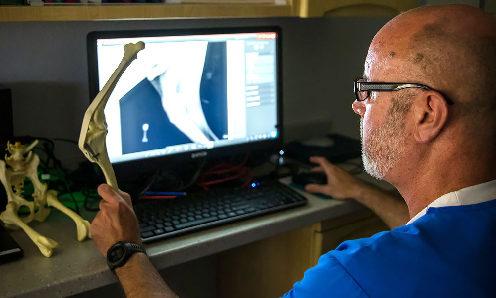
Your veterinarian took radiographs. They were inconclusive. An ultrasound followed and narrowed things down without settling the question. Now someone has said the word CT, and you are wondering whether it will really tell you anything the first two rounds did not.
Usually, yes. And at East Hill Animal Hospital in Pensacola, Florida, you can find out in a few days rather than the few weeks that advanced imaging often requires elsewhere.
Why Layers Beat Shadows
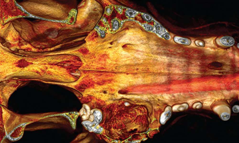
A radiograph flattens a three-dimensional animal onto a two-dimensional image. Everything overlaps. A mass sitting behind a rib, or inside a nasal passage crowded with bone, may simply not be visible. Ultrasound gives excellent soft tissue detail but cannot see through gas or bone, which rules out large portions of the skull and the spine.
CT removes both limitations. It builds a layered view of bone and soft tissue that can be examined section by section.
The Machine Doing the Looking
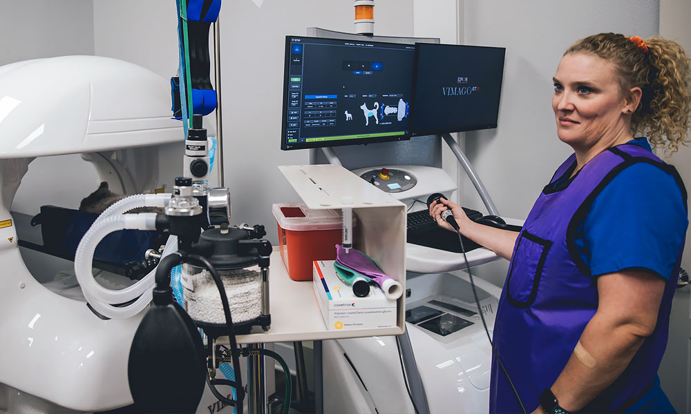
East Hill’s Vimago GT30 goes further than standard CT, and the reason is in what it captures. High-Definition Volumetric Imaging records the anatomy as a volume of data rather than a fixed stack of slices, at a resolution reaching roughly 0.09 millimeters, close to the thickness of a human hair. Afterward the veterinarians can review that volume from any direction and at any slice thickness a given question calls for. Nothing gets locked in at the moment of the scan.
The same system handles fluoroscopy and digital radiology, so a dynamic study can follow a static one without a second appointment.
Every scan is then read by a board-certified radiologist. The equipment produces the detail; a specialist interprets it.
Conditions It Regularly Identifies
Nasal and sinus disease is a common one. Chronic discharge or nosebleeds in a dog can come from a tumor or a fungal infection, and sometimes from something the dog inhaled on a walk. Radiographs of the nasal cavity are notoriously difficult to interpret. CT separates those possibilities.
Brain masses are another. Seizures or circling in an older pet raise the question of an intracranial lesion, and there is no way to answer it with an X-ray.
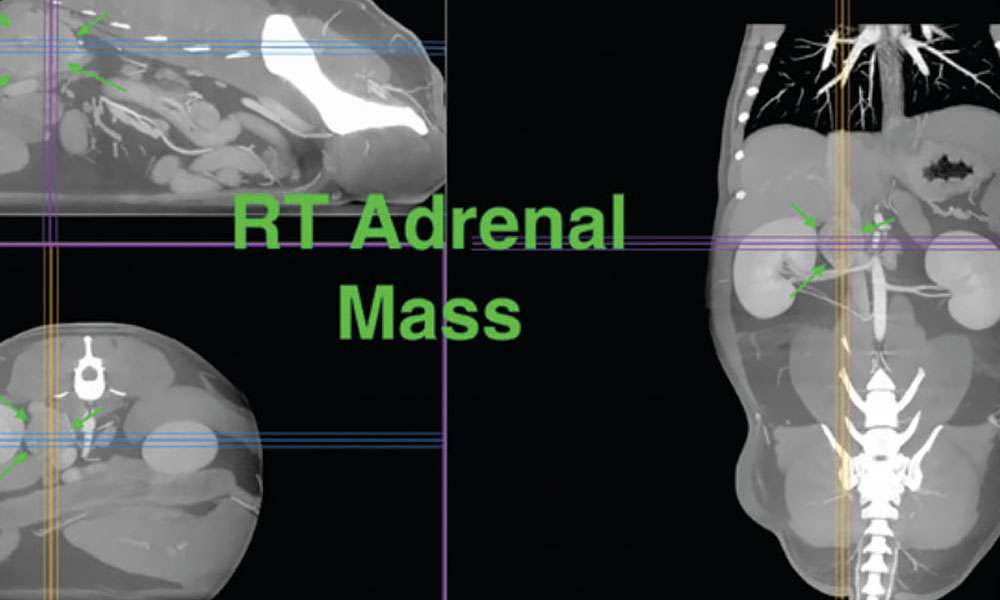
Small abdominal tumors show up as well, including insulinomas in the pancreas that are frequently too small for ultrasound to resolve reliably. The same applies to adrenal disease and to masses in the urethra that hide from conventional imaging.
Upper GI problems belong on this list too. Megaesophagus and hiatal hernia both show clearly on CT, and both are easy to chase for months on a presumptive diagnosis when the imaging is not definitive.
Spinal problems benefit enormously. A dog that suddenly cannot use its back legs needs to know whether the cause is a disc or a fracture, and that answer determines whether surgery is even on the table.
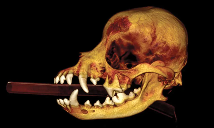
Complex dental disease is a quieter one. Tooth root abscesses and jaw pathology often extend much further than the visible portion suggests, and a jaw fracture running through diseased bone is not something a dental radiograph reliably shows.
Oral masses fall into the same category. Where the tumor stops and healthy tissue begins is a surgical question, and CT answers it better than palpation does.
Orthopedic injuries round out the list. With a complicated fracture, the surgeon needs to know the exact configuration of every fragment before choosing an approach. A 3D reconstruction supplies that in a way flat films cannot.
Why Speed Changes the Answer
None of these conditions pause while you wait for an appointment. A fracture begins healing in whatever position it settled into. A tumor keeps growing, and a spinal injury has a window during which surgery helps most.
Gulf Coast owners have long been sent hours away to Mobile or New Orleans, where the wait is often weeks because those centers serve an enormous region. East Hill schedules CT appointments within days and frequently handles urgent cases the same day, Monday through Friday, with results typically back within days after that.
And What It Costs
Cost stops as many diagnoses as scheduling does. East Hill’s CT pricing generally comes in below other providers in the region, with estimates often running about 75 percent under what a referral institution charges, which means the number is less likely to be the reason a pet goes undiagnosed.
How to Get One Scheduled
Your veterinarian can refer you directly, and East Hill routes results and follow-up communication back to them so your primary vet stays in charge of the treatment plan. You can also call the hospital yourself to ask about availability.
East Hill Animal Hospital is located at 801 E. Cervantes Street in Pensacola and has cared for local pets for over twenty years. Call 850-437-9932 to discuss CT imaging for your dog or cat.